What Can a Dentist Learn from Dental X-Rays That an Exam Cannot Show?

A dental exam can feel reassuring when everything looks healthy. However, a nagging question may remain: could something be hiding beneath the surface? Cavities between teeth, changes around the roots, and bone loss may develop where a mirror and dental instruments cannot reach.
A dentist in Victoria Park uses dental X-rays to view these concealed areas and connect subtle symptoms with their possible causes. Knowing what those images reveal can make the small sensor feel far less mysterious overall.
What Dental X-Rays Add to a Visual Examination
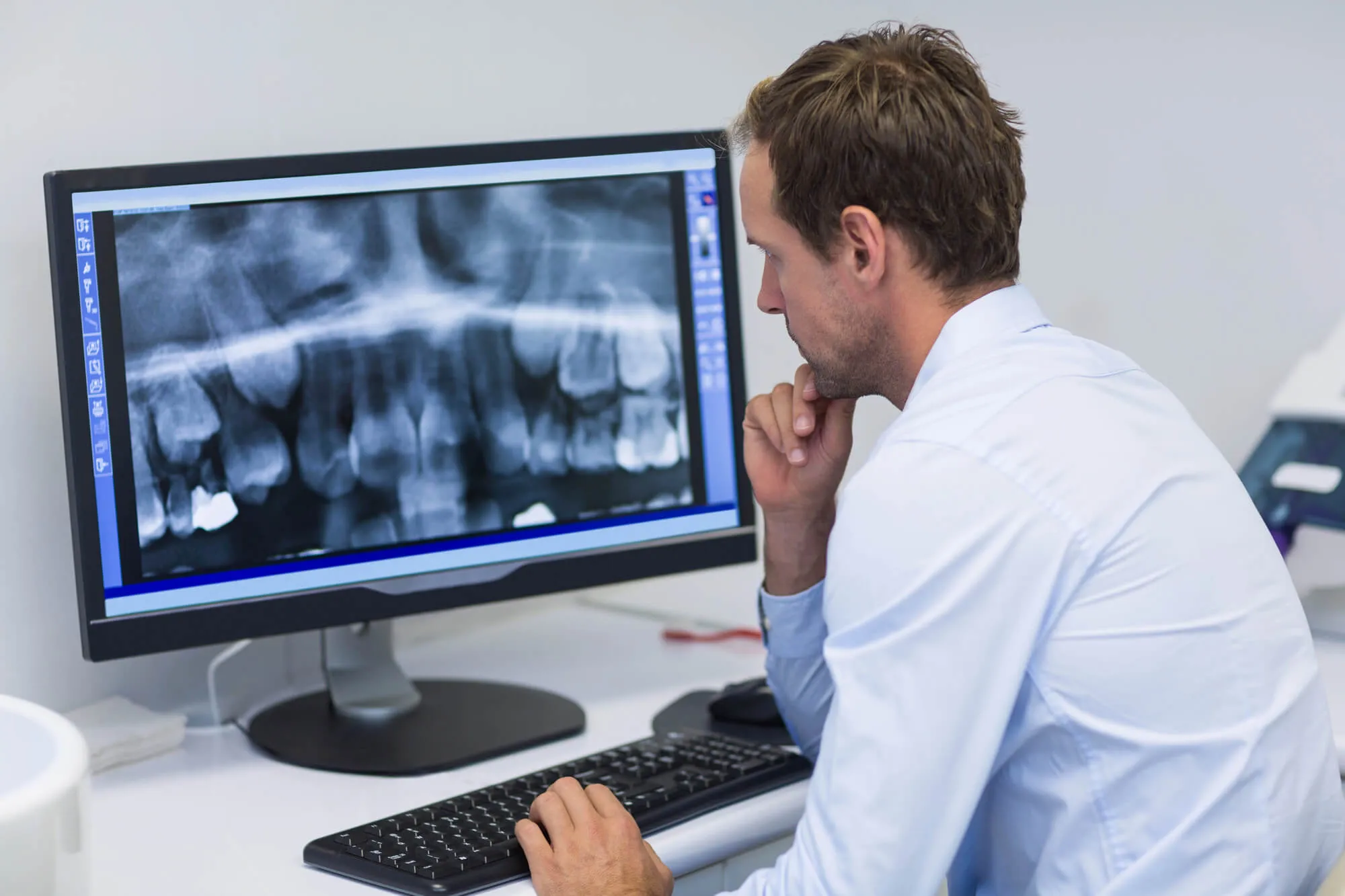
A visual examination allows your dentist to assess your teeth, gums, bite, and existing dental work. X-rays provide another view by showing the roots, supporting bone, and structures below the gums. According to the American Dental Association, they may reveal damage or disease that cannot be seen clinically. Dentists interpret these images alongside your symptoms and health history.
Cavities Between Closely Spaced Teeth
A cavity on the chewing surface of a tooth may become visible during an examination. Decay between two teeth can remain concealed, especially when the teeth sit close together. Even a dental instrument may not reach the affected area.
Bitewing X-rays show the crowns of the upper and lower teeth in a specific section of the mouth. They allow the dentist to examine contact points where food debris and plaque often collect. An image may reveal early changes in enamel or a deeper cavity that has entered the dentin beneath it.
Finding decay at an earlier stage may allow for more conservative care. The dentist can also compare newer images with earlier ones to see whether a suspicious area has changed.
Decay Beneath Fillings and Crowns
Fillings and crowns can remain functional for a long time. However, the natural tooth around or beneath them can still develop decay. This may happen near a restoration’s edge, where bacteria can enter through a small gap.
A crown may look intact from the outside while changes develop beneath it. X-rays can reveal darker areas that suggest decay near a filling, under a crown, or beside another restoration. They may also show whether a filling sits close to the tooth’s pulp, the soft internal tissue containing nerves and blood vessels.
Not every shadow represents a cavity. Dental materials appear differently on radiographs, so interpretation requires knowledge of the restoration, the tooth’s history, and findings from the physical examination.
Infections Around Tooth Roots
The portion of a tooth visible above the gums represents only part of its structure. Each tooth also has one or more roots anchored within the jawbone. Problems around these roots may cause pain, pressure, swelling, or sensitivity, but some produce few noticeable symptoms.
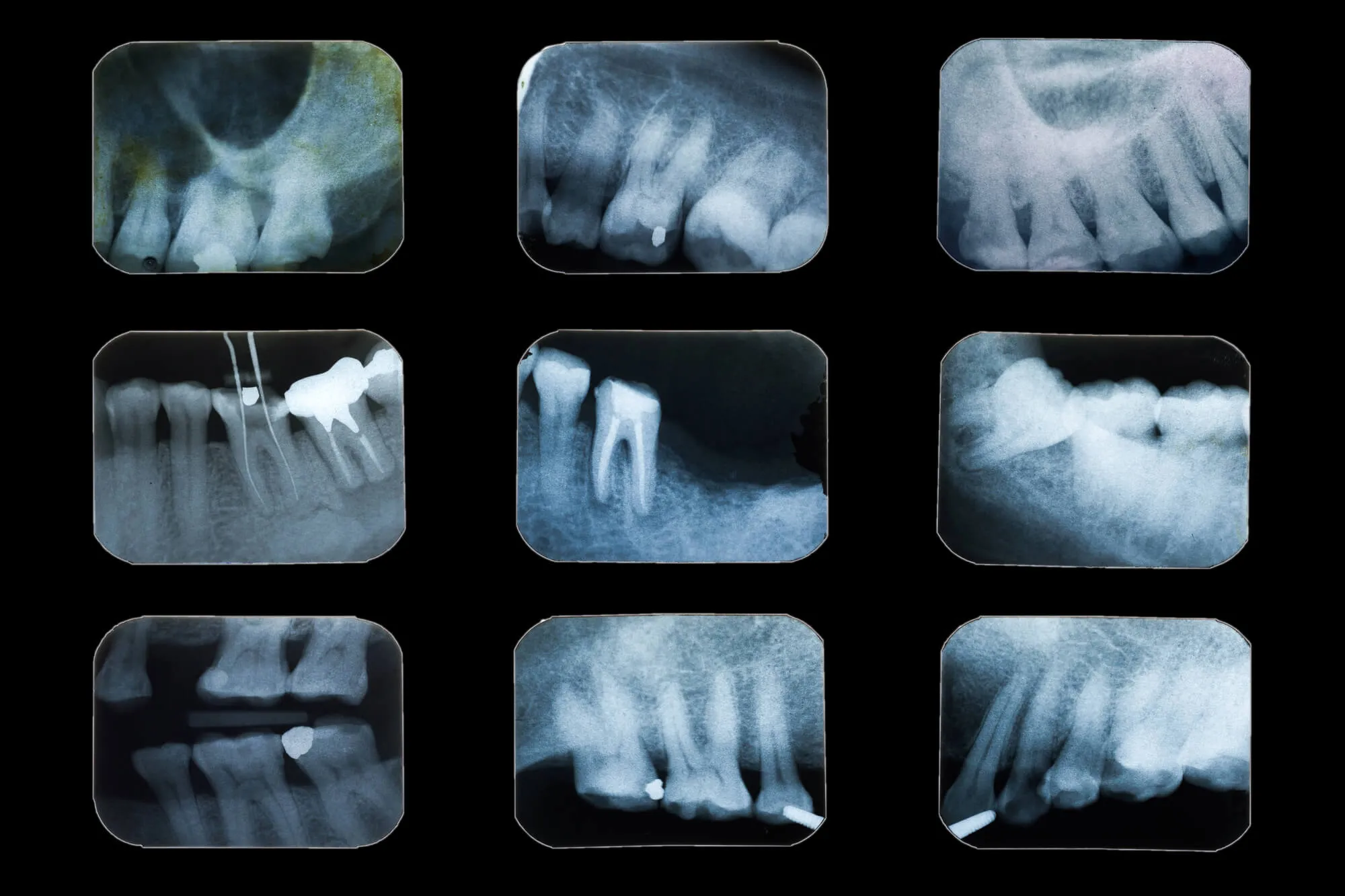
A periapical X-ray captures the entire tooth, from the crown to the root tip. It can reveal bone changes near the root tip that may be associated with infection or inflammation. It may also show the number, shape, and direction of the roots.
This information can support planning for root canal treatment or extraction. After a root canal procedure, radiographs may help the dentist evaluate the filled canals and monitor the bone surrounding the tooth.
Bone Loss Around the Teeth
Gums can appear mildly irritated even when bigger changes have begun around the supporting bone. Since bone lies below the gumline, the dentist cannot assess its condition solely by visual inspection.
X-rays help show the height and shape of the bone around each tooth. A reduction in bone level may indicate periodontal disease, particularly when it occurs alongside gum pocket measurements, bleeding, recession, or tooth mobility.
Radiographs can also reveal whether bone loss affects a single area or multiple areas of the mouth. That pattern helps the dental team assess the extent of the condition and plan appropriate periodontal care. Future images may be compared with earlier records to monitor further changes.
Impacted Teeth and Hidden Tooth Development
A tooth is considered impacted when it cannot erupt into its expected position. Wisdom teeth commonly become trapped beneath the gums or jawbone, but other teeth can also be affected.
A panoramic X-ray provides a broad image of the upper and lower jaws. It can show the position and angle of impacted teeth, their proximity to neighboring roots, and nearby anatomical structures. These details help the dentist determine whether observation or oral surgery is appropriate.
In younger patients, X-rays can show developing adult teeth that have not erupted. They may reveal missing teeth, extra teeth, crowding, delayed development, or unusual eruption paths that are not visible during an examination.
Changes Within the Jawbone
Dental radiographs sometimes reveal cysts, unusual bone patterns, fractures, or other changes in the jaw. A visual examination may provide clues, such as swelling or altered sensation. Still, it cannot show the full area beneath the tissue.
An unusual image does not automatically confirm a diagnosis. The dentist may recommend another type of image, monitor the area, or arrange an evaluation by a dental or medical professional with relevant training.
Dental X-rays also have limits. Very fine cracks, early soft-tissue changes, and some sources of pain may not be clearly visible. A normal-looking radiograph does not dismiss a patient’s symptoms. It becomes one part of a broader assessment.
Planning Dental Implants and Restorative Care
Images provide measurements and anatomical details that can support planning for dental implants, crowns, bridges, dentures, and other restorative services. For implant care, the dentist needs information about the jawbone and the location of nearby structures.
Standard two-dimensional images may provide enough information for some situations. A cone-beam computed tomography (CBCT) scan provides three-dimensional views when greater detail is clinically necessary. CBCT may help assess bone dimensions, implant position, impacted teeth, or complex root anatomy.
The ADA’s updated radiography recommendations state that dental imaging should be ordered only when clinically necessary. The type of image depends on the patient’s symptoms, oral health, disease risk, age, history, and planned dental care.
How Often Are Dental X-Rays Needed?
Dental X-rays do not follow one schedule for everyone. Your dentist considers your symptoms, oral health, dental history, and risk of disease before recommending them. Recent images from another practice may also be useful. Modern digital systems use low radiation levels, while careful image selection and positioning limit exposure to the areas that need evaluation.

See Beyond the Surface with a Dentist in Victoria Park
Dental X-rays can reveal decay, infection, bone loss, and developing teeth that a visual examination cannot show. At 1500 Dental, our team combines patient education, compassionate care, and modern technology to support your dental and overall health. Contact us to schedule an appointment and receive care planned around your needs.